Most mouth ulcers and sores are nothing to worry about. They come from a knock, a sharp tooth, a hot drink or a run-down week, and they settle on their own within about two weeks. The one to pay attention to is different. A mouth sore that won’t heal after two to three weeks, or a lump, or a patch that simply stays, deserves a proper look rather than more waiting.
This is not a reason to panic. The large majority of these changes turn out to be harmless. The point of getting them checked early is simple: on the rare occasion that something is serious, finding it early makes it far easier to treat. This guide explains which signs are worth acting on, what white and red patches can mean, and when a small procedure called a biopsy is the sensible next step.
Most Mouth Sores Are Harmless, but Some Are Not
An ordinary ulcer has a clear cause and a clear end. It hurts for a few days, then fades and heals within roughly two weeks. Because that pattern is so reliable, doctors and dentists use it as a rule of thumb. Anything in the mouth that has not healed after two to three weeks has outstayed its welcome and should be examined.
The reassuring truth is that unhealed sores are common and usually benign. A denture rubbing, a rough filling, a cheek you keep biting, or a viral infection can all keep a sore going. A dentist can often spot and remove the cause on the spot. The reason the two-week rule still matters is that the small number of sores that are not routine tend to look ordinary at first, so time is the clue that separates them.
Common Reasons a Sore Lingers
Before worrying, it helps to know how often there is a simple explanation. A great many stubborn sores trace back to ongoing irritation that keeps reopening the same spot. A denture that no longer fits, a chipped or sharp tooth, an old filling with a rough edge, or a habit of biting the same piece of cheek can all keep a sore going for weeks. Remove the cause and the sore usually heals as expected.
Infections and general health play a part too. Cold sores from a virus, fungal infections such as oral thrush, and flare-ups of conditions like lichen planus can each produce sores or patches that come and go. Nutritional gaps, certain medicines, stress and a run of poor sleep can slow healing as well. A dentist can tell many of these apart by sight and history, which is another reason a persistent sore is better examined than watched at home. Naming the cause is often all the reassurance a person needs.
Warning Signs Worth a Closer Look
You do not need to memorise a medical list. A few plain signals are enough to prompt a visit.
- A sore, ulcer or raw area anywhere in the mouth or on the lip that has not healed in two to three weeks.
- A lump, swelling or thickening in the cheek, gum, tongue or floor of the mouth that you can feel with your tongue or finger.
- A white patch that will not wipe or brush away.
- A red patch, or a mixed red and white patch, that stays.
- An area that bleeds easily without an obvious knock.
- Numbness, persistent pain, or a burning feeling that does not settle.
- Trouble chewing, swallowing or moving the tongue or jaw, or a feeling that something is caught.
- A tooth that becomes loose for no clear dental reason.
- A one-sided sore throat, hoarseness or ear pain that lingers.
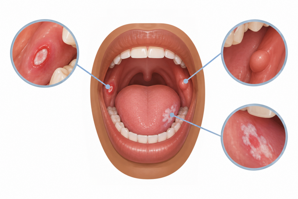
Any single one of these is usually harmless on its own. What raises the priority is persistence. If it lasts, get it seen.
What White and Red Patches Can Mean
Some patches in the mouth belong to a group that dentists call oral potentially malignant disorders. The name sounds heavy, but it simply means these areas carry a slightly higher chance of change over time and are worth watching or testing, not that they are cancer.
A white patch that cannot be rubbed off is known as leukoplakia. Most never cause harm. Pooled research across many studies puts the average chance of a leukoplakia turning into something more serious at roughly seven percent, and that risk is higher when the patch is on the side of the tongue, is mixed in appearance rather than uniform, or occurs in someone who smokes.³ Those details are exactly why a dentist wants to see it rather than guess.
A red patch, called erythroplakia, is less common but more important. Among these lesions it carries the highest risk of change, which is why any persistent red or speckled red and white area should be examined promptly rather than watched from home.⁴ Redness in the mouth can have many innocent causes, so the aim is not alarm but a clear answer.
Why Getting It Checked Early Matters
Mouth cancers are far more treatable when they are found early and small. The difficulty, across many countries, is that they are often found late, after the change has had time to grow. Reviews of the evidence describe this as one of the main challenges in the field, and they point to missed or delayed recognition as a key reason outcomes lag behind what they could be.²
Delay is not only a patient issue. A recent analysis found that the time between a first visit to a health professional and a referral to a specialist accounts for about a third of the whole journey to diagnosis.¹ In plain terms, both halves matter: acting on a sign quickly, and being seen by someone who examines the mouth carefully and refers on when needed. Choosing a dental visit for a persistent mouth change puts you in front of exactly the kind of examination that is built for spotting these things.
The reason timing matters so much comes down to how these conditions behave. A small, early change is usually easier to treat, needs less involved treatment, and carries a better outlook than the same problem found months later once it has grown or spread. That gap between early and late is the whole argument for the two-week rule. It costs very little to be checked, and the peace of mind of a clear result is worth the short visit on its own. Regular dental visits add another layer, since a dentist looking in your mouth routinely is well placed to notice a change you might not, sometimes before it causes any symptom at all.
What Puts Someone at Higher Risk
Certain habits and factors raise the background risk, and knowing them helps you judge when to be a little more watchful.
- Tobacco in any form, whether smoked or the chewed and placed types.
- Alcohol, especially heavy use, and more so when combined with tobacco.
- Areca nut and betel quid, common in some communities.
- Long term sun exposure, which mainly affects the lips.
- Older age, and a previous mouth lesion or cancer.
It is worth saying clearly that mouth cancer can also appear in people with none of these factors, and that many people with these habits never develop it. Risk factors raise the odds, they do not decide the outcome, so a persistent sign is worth checking whatever your history.
The practical takeaway is that reducing these habits helps on two fronts at once. Cutting down or stopping tobacco, alcohol and areca nut lowers the long term risk, and it also helps ordinary sores heal faster, since these habits irritate the lining of the mouth and slow repair. Protecting your lips from strong sun with a barrier balm is a small, easy step for anyone who spends long hours outdoors. None of this replaces getting a lasting change looked at, but it does tilt the odds in your favour over time.
How a Diagnosis Is Made, and When a Biopsy Is Needed
The first step is always a careful examination. A dentist looks at every surface of the mouth and lips, feels for lumps in the cheeks, tongue, floor of the mouth and neck, and asks about how long the change has been there and how it has behaved. The soft tissue check is quick and painless, and it covers areas that are easy to overlook at home, such as the underside of the tongue and the floor of the mouth. Bringing a simple note of when you first noticed the change, and whether it has grown, bled or altered in colour, makes this part faster and more useful.
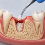
If an area looks routine and has an obvious cause, the dentist may simply remove the cause and review it in a couple of weeks to confirm it heals. If an area is suspicious or has not settled, the definitive step is a biopsy. This means taking a small sample of the tissue so it can be examined under a microscope in a laboratory. A biopsy combined with the clinical examination remains the reliable standard for a firm diagnosis, and the various light or dye based screening gadgets that exist are helpful extras at most, not replacements for it.⁵
The area to be sampled is numbed first, so the procedure itself is not painful, and the small site usually heals within a week or so with simple aftercare. A biopsy is a minor procedure, usually done under local anaesthetic, and it is the only way to know for certain what an area is. It is also worth holding on to the reassuring side of this: many biopsies come back clear or show a harmless cause, and even when they find a potentially malignant patch, catching it at that stage is precisely the early window that makes management far more straightforward.
What to Do If You Notice Something
The sensible middle path is neither to panic nor to wait and hope. If a sore, lump or patch in your mouth has lasted more than two to three weeks, book a dental examination. Note when you first saw it and whether it has changed, since that history helps the dentist. Avoid poking or repeatedly testing the area, and if you use tobacco or areca nut, cutting back or stopping helps both the healing of ordinary sores and your longer term risk.
It also helps to resist the temptation to self-diagnose from photos online, since many harmless and serious changes can look alike to an untrained eye, and the comparison usually adds worry rather than clarity. If the sore is painful in the meantime, a gentle salt water rinse and avoiding sharp, spicy or very hot foods can keep it comfortable while you wait for your appointment. Bring a list of any medicines you take and any habits worth mentioning, as both can shape what the dentist looks for.
A single visit often settles the question entirely, either by finding a simple cause or by arranging the right next step without delay.
Caring for Your Smile at DentMind CBD
A mouth change that will not settle is worth an unhurried, expert look rather than weeks of quiet worry. If you have a sore, lump, or white or red patch that has lasted more than a couple of weeks, the team at DentMind CBD Dental Centre on Kimathi Street in Nairobi CBD can examine it properly, explain what they find in plain terms, and arrange any further step needed. You can book an appointment online, send a quick question on WhatsApp , or read more across our blog at dentmindcbd.co.ke, and give yourself the reassurance of a clear answer sooner rather than later.
This article is general information and NOT a diagnosis. Any persistent change in your mouth should be assessed by a dental or medical professional.
Footnotes
- Seoane Romero, J. M., Mallah, N., Varela-Centelles, P. I., Warnakulasuriya, S., & Takkouche, B. (2025). Primary care referral delays in oral cancer diagnosis: a meta-analysis. Oral Diseases, 27(7), 2148–2159. https://doi.org/10.1111/odi.15218
- Gonzalez-Moles, M. A., Aguilar-Ruiz, M., & Ramos-Garcia, P. (2022). Challenges in the early diagnosis of oral cancer, evidence gaps and strategies for improvement: a scoping review of systematic reviews. Cancers, 14(19), 4967. https://doi.org/10.3390/cancers14194967
- Guan, J. Y., Luo, Y. H., Lin, Y. Y., Wu, Z. Y., Ye, J. Y., Xie, S. M., & Li, J. (2023). Malignant transformation rate of oral leukoplakia in the past 20 years: a systematic review and meta-analysis. Journal of Oral Pathology & Medicine, 52(8), 691–700. https://doi.org/10.1111/jop.13440
- Lorenzo-Pouso, A. I., Lafuente-Ibáñez de Mendoza, I., Pérez-Sayáns, M., Pérez-Jardón, A., Chamorro-Petronacci, C. M., Blanco-Carrión, A., & Aguirre-Urízar, J. M. (2022). Critical update, systematic review, and meta-analysis of oral erythroplakia as an oral potentially malignant disorder. Journal of Oral Pathology & Medicine, 51(7), 585–593. https://doi.org/10.1111/jop.13304
- Lau, J., O, G., Warnakulasuriya, S., Balasubramaniam, R., Frydrych, A., & Kujan, O. (2024). Adjunctive aids for the detection of oral squamous cell carcinoma and oral potentially malignant disorders: a systematic review of systematic reviews. Japanese Dental Science Review, 60, 53–72. https://doi.org/10.1016/j.jdsr.2023.12.004